
Specialist ultrasound
Ultrasound assessment of the female pelvis, with a focus on endometriosis mapping.
Learn about endometriosis mappingSpecialist radiology • São Paulo
Specialist ultrasound, MRI second opinions, a neuropelveology protocol and MR angiography assessment.

Imaging exams and reports
Explore the areas of practice and choose what you would like to discuss with the team.
Ultrasound assessment of the female pelvis, with a focus on endometriosis mapping.
Learn about endometriosis mappingA new specialist review of images from a previously performed MRI, subject to an assessment of whether the material is suitable.
Learn about the second-opinion serviceMRI assessment directed at the pelvic nerves and related structures, according to the indication.
Learn about this assessmentSpecialist assessment of MR angiography exams, subject to the indication and technical adequacy of the material.
Learn about this assessmentSpecialist ultrasound
A dedicated imaging assessment for the investigation of endometriosis, taking into account the available clinical information and the purpose of the exam.
Contact the team to check availability and receive instructions for the exam.
MRI second-opinion report
A second-opinion report provides a new specialist review of your exam images. Image quality and suitability are assessed before the review can be confirmed.
Tell the team that you are requesting an MRI second-opinion report.
Receive instructions for securely providing the images and required information.
Image quality and suitability are checked before the service is confirmed.
Once confirmed, the team will explain the report process and delivery.
When the images are suitable, the review may be performed using the existing exam. The team will explain the next steps.
Specialist magnetic resonance imaging
The scope of practice also includes MRI with a neuropelveology protocol and MR angiography assessment. Indication, availability and technical adequacy are reviewed individually by the team.
A protocol directed at imaging the pelvic nerves and related structures according to the clinical question.
MRI assessment focused on vascular structures according to the region and clinical indication.
Information about indication, documents, availability, fees and turnaround time is provided exclusively through WhatsApp.
About the radiologist
Cinthia Callegari Barbisan is a radiologist whose practice is dedicated to female pelvic imaging and endometriosis. She completed her radiology residency and fellowship at BP — A Beneficência Portuguesa de São Paulo.
Her work combines diagnostic imaging, teaching and scientific activities. Each exam is interpreted in context, with attention to the available clinical information, careful image review and communication with the referring physician.

Teaching and research
Participation in medical education, conferences and scientific work focused on women’s imaging.
Training radiologists and contributing to continuing medical education.
Lectures and participation in national and international conferences on female pelvic imaging and endometriosis.
Author and co-author of publications and scientific work in women’s imaging.
Patient-friendly radiology information: what each exam can show and how imaging contributes to the investigation of endometriosis.
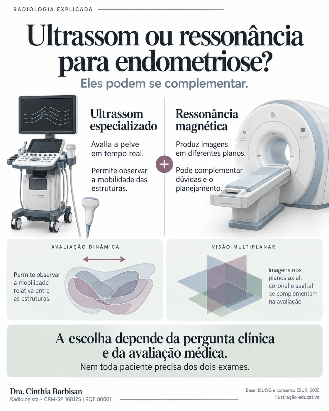
The methods may complement each other. The choice depends on the clinical question the exam needs to address.
Specialist ultrasound allows real-time assessment of the pelvis, including the mobility of pelvic structures. Magnetic resonance imaging (MRI) produces images in different planes and may complement the investigation or treatment planning.
Not every patient needs both exams. The choice considers symptoms, previous results and the purpose of the assessment.
Reference: ESUR consensus MRI for endometriosis: indications, reporting, and classifications (2025)
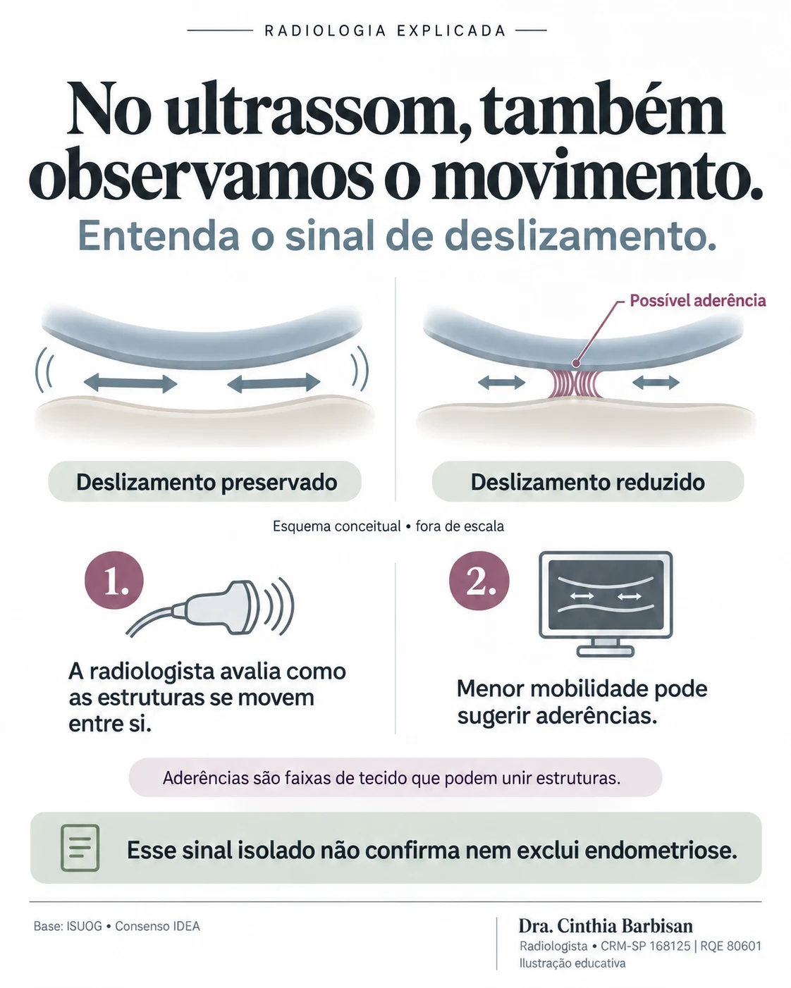
Learn about the sliding sign and how it helps assess the mobility of pelvic structures.
During ultrasound, the radiologist can observe how structures slide in relation to one another. This movement is part of the dynamic assessment.
Reduced or absent sliding may be associated with adhesions, which are bands of tissue that connect structures. This sign is interpreted together with the other findings and clinical history. On its own, it neither confirms nor rules out endometriosis.
Reference: ISUOG VISUOG — Bowel endometriosis / avaliação dinâmica e sinal de deslizamento
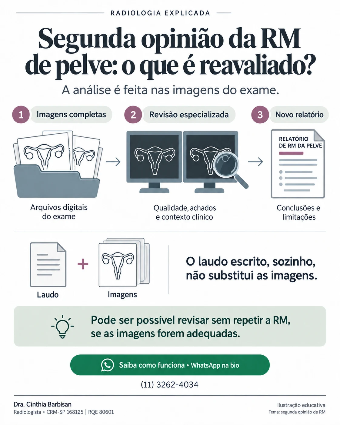
Learn why the complete images, exam quality and clinical information matter.
A second opinion is a new review of the images from a previously performed MRI. Technical quality, imaging findings and the available clinical information are considered. The written report is helpful, but it does not replace the complete image set.
If the images are suitable, the review may be possible without repeating the MRI. The new report presents the conclusions and limitations of the assessment.
Reference: RadiologyInfo — Your Medical Images
Information about MRI second opinionsConfirm the requested exam, location, preparation and required documents. Follow the specific instructions provided by the team for your exam.
State whether you are seeking specialist ultrasound for endometriosis mapping or an MRI second-opinion report. For an image review, explain that the exam has already been performed and wait for instructions on the appropriate channel for providing images and documents.
Educational content; it does not replace an individual medical assessment. Illustrations are conceptual and do not represent patient exams.
Frequently asked questions
MRI second opinions, specialist ultrasound for endometriosis mapping, a neuropelveology protocol and MR angiography assessment.
The existing images are assessed first to determine whether they are suitable for review. The team will confirm suitability and explain the next steps.
Contact the team on WhatsApp for instructions on providing images and the required information.
Indication, availability, preparation, documents, location, fees and turnaround time are provided exclusively by the team through WhatsApp.
Imaging exams and reports
WhatsApp is the exclusive channel for information about imaging exams, reports and specialist protocols.
Indication, preparation, documents, location, availability, fees and turnaround time are provided through WhatsApp.